HIV Foundation Health Biliary cancer often causes no symptoms – and is therefore usually recognized too late

Biliary cancer often causes no symptoms – and is therefore usually recognized too late

Gallbladder cancer in particular only leads to symptoms in an advanced stage. Why this is so, what role gallstones play – and why the prognosis so far has often been unfavorable.

Biliary cancer, with around 5500 new cases per year, is one of the rare forms of cancer, but it is particularly risky. According to popular opinion, the tumor causes almost no early symptoms and is therefore usually only recognized late when an operation is no longer possible and the tumor has already metastasized.

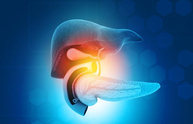
Biliary cancer – important: inside or outside the liver

The fact is, however, that the colloquial term biliary cancer, medically cholangiocarcinoma (CCA), covers different forms. First of all, there is gallbladder cancer, which forms in the gallbladder, which in turn is embedded in the liver.

Secondly, a carcinoma can form in the bile ducts, which are not only located within the liver and direct the bile to storage in the gallbladder, but also away from the gallbladder, which lead the bile to the small intestine.

“Depending on the localization, we differentiate between intra- and extrahepatic carcinoma, i.e. those that develop inside or outside the liver,” explains Arndt Vogel, spokesman for the “Hepatobiliary Tumors” working group of the Internal Oncology Working Group (AIO) and head of the Visceral Oncological Center Hannover Medical School (MHH).

The risk of developing cholangiocarcinoma increases with age. Overall, the incidence of intrahepatic carcinomas is increasing, while that of extrahepatic carcinomas falls somewhat.

Risk factors for biliary cancer

An exception in connection with cholangiocarcinoma is Southeast Asia, especially countries like Thailand. This cancer often occurs there because certain parasites can inflame the biliary tract. Chronic inflammation plays an important role in the development of biliary cancer.

The following risk factors come into play in the western industrialized nations, but they are also closely related to inflammation:

  • Primary sclerosing cholangitis, an inflammation of the bile ducts that mostly affects men.
  • Cysts in the bile and bile ducts, including Caroli’s syndrome; they increase the risk of biliary cancer.
  • Smoking, because the substances in smoke are known to be carcinogenic, are not only excreted via the kidneys and urine, but are also collected, processed and passed on in the bile.
  • Gallstones; However, only when they cause problems, i.e. inflame the bile, do they promote the development of cancer.

Gallstones and biliary cancer

Around ten percent of Germans are said to have gallstones, and the risk increases with age. “But very few of those affected develop cholangiocarcinoma. This cancer is very rare, ”says the medicine professor reassuringly.

The gallbladder should only be removed if the stones cause problems, i.e. colic and inflammation.

Symptoms appear differently late, but are similar

The signs of gallbladder inflammation caused by stones are somewhat similar to those of cholangiocarcinoma (CCA).

So biliary cancer can cause the following signs:

  • Jaundice (jaundice)
  • nausea
  • Vomit
  • Pain in the left upper abdomen.

The location of the carcinoma is crucial for the stage at which symptoms appear:

  • Intrahepatic carcinoma triggers these clear signs quite late, “because the liver doesn’t hurt when a tumor grows there,” explains the expert.
  • Extrahepatic carcinoma, on the other hand, usually quickly means that the bile can no longer flow into the intestine. Bile congestion and jaundice are relatively early signs of this type of cancer.

That is why bile duct cancer that grows outside the liver is usually diagnosed earlier – but it is difficult to operate because of its often complicated location next to blood vessels and does not make the generally difficult situation with cholangiocarcinoma any easier, the oncologist limits the associated high expectations.

Diagnosis of cancer of the gallbladder and bile ducts

Doctors use cross-sectional image diagnostics such as MRI and CT. “This allows the suspicion to be clarified and the staging, i.e. stage and spread, to be identified,” explains Vogel.

The histological examination provides additional details about the tumor, whereby the samples in gallbladder cancer are relatively easy to obtain. However, this is more difficult with extrahepatic tumors because the biliary tract is often narrow and winding. The examination is carried out through an endoscope, the method here is called endoscopic retrograde cholangiopancreatography (ERCP examination).

Are there any less invasive methods? Ultrasound, carried out endoscopically through the stomach from the inside or from the outside, can also be informative, says the cancer specialist. However, the methods of first choice are MRI and CT.

Treatment of biliary cancer – surgery not always possible

If the suspicion has been confirmed and the results of the examination enable the tumor to be classified, the goal is to remove the carcinoma surgically. “However, as already described, this is sometimes difficult due to the location of the tumors,” reports Vogel. However, the surgical techniques have improved significantly in recent years.

The standard treatment for patients with advanced tumors is chemotherapy, with a combination of gemcitabine and cisplatin.

In a palliative situation, i.e. to lengthen survival time and / or improve quality of life, local therapies such as selective internal radiotherapy (SIRT, radioembolization) are currently used in clinical studies . Radioactive microspheres are guided to the tumor via an inguinal catheter, its cells are destroyed and healthy tissue is spared. The first results show that for some patients many months can be gained with this.

The prognosis for biliary cancer is poor …

Despite all these possibilities, few patients can be cured. Even if the tumor could be completely removed in the healthy, the recurrence rate is still relatively high. “60 to 80 percent of the tumors come back,” reports Vogel. Because the tumors spread very early.

… but with the therapy “a small revolution is emerging”

This is the bad news. In fact, these prospects could improve in the future. The oncologist says: “Because a small revolution is taking place here at the moment.” The interest of pharmaceutical companies in this rare cancer has increased significantly, and intensive work is being carried out on the development of new drugs.

The reason for this change is the fact that it has been discovered that numerous genetic changes occur in these tumors and thus allow a molecular, i.e. targeted therapy. There have been many studies on this topic for a few years now.

Two developments are particularly promising:

1. Inhibitors against IDH1 mutations , from which patients with a corresponding cholangiocarcinoma can clearly benefit.

2. Inhibitors against FGFR2 , fusions, MSI, NTRK and others.

“There are currently a number of very promising active ingredients in the test that have the various genetic changes as a starting point,” reports the oncologist. How much these new therapies could improve the treatment of biliary cancer becomes clear when one realizes that 40 to 50 percent of all these tumors, especially intrahepatic ones, show such genetic changes and are therefore suitable for targeted, molecular therapy.

Prevention Of Bile Cancer – Quit Smoking!

However, it will be some time before the new therapies are available to all patients. Until then, it is still true that biliary cancer is difficult to treat and the prognosis is unfavorable.

This makes prevention all the more important. To what extent can everyone prevent this tumor – apart from the advice not to smoke, which is so important with regard to many other diseases? The expert also has one recommendation in particular:

Get gallstones cleared up if they’re causing problems. However, this does not mean that everyone who has gallstones should be afraid: Gallstones are considered to be risk factors for gallbladder cancer, but only one percent of all gallstone carriers develop this tumor.

Leave a Reply

Your email address will not be published. Required fields are marked *

Related Post

Key Principles of Implant DentistryKey Principles of Implant Dentistry

Before your consultation with your dentist at implant dentistry san diego, you should know what to expect from your procedure. The main points of this article are the importance of soft tissue, bone, and implant placement. Occlusal contact is also a crucial point to consider. All these points should be carefully examined before treatment. The goal of your GP is to protect you and the staff. Before you go in for your appointment, ask yourself if you have any of these risk factors.

Bone

Whether the procedure is a flap-less or traditional surgery, it requires careful planning. In either case, a series of drills gradually enlarges the site of the implant. The final bur is slightly smaller than the implant itself, and it is placed with a torque-controlled wrench to avoid overheating the bone. The bone surrounding the implant is then reshaped to accommodate the new fixture.What is a Dental Implant Same Day Procedure? - Smiles By Julia Fort  Lauderdale Florida

Osteointegration occurs when the biomaterial that is used to support dental implants is osteoconductive. This material enables the dental implant to integrate with the bone surface. Histological studies have shown that implant surface contacts the host bone and initiates the healing process. The procedure follows a sequence similar to that of bone regeneration in fractures and small defects. It ends with “restoration ad integrum,” or the absence of scar tissue.

Soft tissue

The success of dental implants depends on the stability of soft tissue surrounding the implants. This stability is best achieved through proper diagnosis and surgical planning. Inadequate soft tissue evaluation can lead to improper placement of dental implants, which is detrimental to both patient and implant health. Soft tissue grafting can be performed to address these soft tissue concerns. Grafting techniques differ according to patient anatomy and morphology. The biotype of the gingival tissues is based on the morphology of the tooth, bone, and gum tissue. An underdeveloped biotype can lead to pocket formation. A biotype with thick, flat tissue may not be suitable for implant placement.

The graft site must provide adequate vascularisation for the graft. It must be rigidly immobilised and provide adequate hemostasis. The donor tissue must be large enough to facilitate immobilization and achieve the desired volume augmentation after secondary contraction. Soft tissue grafting should be performed before the bone grafting procedure. If soft tissue grafting is not possible, connective tissue autografts can be used.

Implant placement

If successful dental implant placement is the ultimate goal, then a modern dentist will implement reproducible treatment protocols, which will ultimately lead to more successful outcomes. There are five key principles of implant dentistry, including past medical history, examination, occlusion, dental imaging, fixed versus removable prosthodontics, and surgery. This article will discuss each of these concepts and their proven contributions to implant dentistry. You should consider implementing these principles into your practice, too.

While successful implant placement is crucial, it’s also the most challenging aspect. Implant placement can go wrong, causing the implant to misalign with the bone. This can lead to unnatural crowns, gum recession, and dark grey metal around the gum. Improper implant placement can result in poor oral health and failure. In order to minimize these risks, patients should follow oral hygiene and care instructions closely for the long-term success of their implants.

Occlusal contact

Occlusal contact is an important component of implant prosthesis and must be accounted for. Ideally, occlusal contact should occur over a flat surface perpendicular to the implant body and be centered over the implant abutment. Secondary occlusal contacts should be placed within one millimeter of the implant body’s periphery to reduce the moment loads. Contacts with the marginal ridges should be avoided, as they are the most susceptible to cantilever forces and should be recontoured to occlude with the central fossa.

When there is an occlusal disparity, the dentist can utilize articulating papers or ribbons to check the occlusion. While these devices leave a contact mark, they are not effective because they are too thick and don’t give enough information to detect an occlusal disparity. Further, the thicknesses of dental marking ribbons vary from 20 microns to 200 microns.

Maintenance

Among other things, the maintenance phase of dental implants entails various parameters and risk factors. During this phase, patients should be informed of the procedures and associated risks. This is because a maintenance procedure involves the continued replacement of a dental implant with a new one. Informed consent is the key to a successful maintenance process. Here are some of the most critical aspects of the maintenance phase. Read on to learn more about the important aspects of this phase of dental implants.

A typical dental implant maintenance visit should last approximately an hour. The dentist will check for any changes to the patient’s medical and dental history. Instrument selection is also important, to avoid trauma to the implant surface or peri-implant tissues. Although an increasing number of patients opt for dental implants, they should understand the challenges of their treatment and follow their recommendations for routine dental care. Listed below are some tips for maintaining dental implants:

Complications

There are a number of complications associated with dental implants. While these complications are relatively rare, they can still occur. Listed below are some of the most common ones. These complications can occur for several reasons, including bone loss, smoking, gum disease, or poor oral hygiene. If you experience any of these symptoms after implant dentistry, contact your dentist as soon as possible. If you’re unsure whether implant surgery is right for you, read on to learn about the potential complications and how to prevent them.

Biomechanical stress is the leading cause of implant failure. This stress causes the implant to fail early or fracture, abutment or prosthetic screw to loosen, and implant crestal bone to collapse. Additionally, implants can become loose and cause overdentures to fall out. These complications are avoidable through a systematic engineering approach, including the identification of underlying causes. In general, implant placement is a successful procedure in 95% of cases.

Osteoarthritis in the knee: How stem cells can repair damaged cartilageOsteoarthritis in the knee: How stem cells can repair damaged cartilage

It crunches and cracks, and some movements during exercise are really painful. When the knee shows signs of wear and tear, those affected want a miracle cure that rebuilds the cartilage. Hope rests on stem cells.

  • The body’s own stem cells have a positive effect on osteoarthritis-related inflammation in the knee.
  • A study now wants to clarify whether they actually build up permanent cartilage.
  • A transplant can repair minor cartilage damage in young patients.

After a certain age, signs of wear and tear become noticeable in the knee . After the age of 30, the risk increases linearly. The painful, inflammatory breakdown of cartilage in the operating room and with an artificial knee ends 150,000 times a year. Then smooth metal has to replace the cartilage layer, which allows round, painless movements in a healthy knee.

The idea frightens many people suffering from osteoarthritis. They hope for new methods to rebuild lost cartilage: stem cells should help.

Belly fat provides the best stem cell material

The doctor uses the patient’s belly fat as a starting material. The idea behind it: stem cells can be obtained particularly easily and in relatively large numbers from vascular fat tissue. Injected at the location of the defect, they learn from the microenvironment into which cell type they should develop.

According to this principle, the doctor and stem cell researcher Eckhard Alt uses the undifferentiated cells : Stem cells from the patient’s fat tissue are processed in the operating room within an hour and injected into the patient where he needs them – for example into the osteoarthritis knee.

The founder of the “Interdisciplinary Stem Cells Research Center” in Houston and a private clinic in Munich sees stem cells as the future therapy for chronic inflammatory diseases of the musculoskeletal system – among other things.

The cell extract in the knee does not guarantee success

Some orthopedic practices that offer the procedure honestly state that it is a not generally recognized attempt at healing with no guarantee of success. Rather, it is a final experimental attempt to remedy knee problems without a joint replacement.

“In this so-called ‘point-of-care’ application, a cell extract is injected that not only consists of stem cells,” explains Oliver Pullig from the Fraunhofer Translational Center for Regenerative Medicine in Würzburg. How many stem cells that are supposed to develop into cartilage material actually get into the knee is just as little regulated as the preparation of the suctioned off belly fat.

Development of osteoarthritis

Osteoarthritis most commonly occurs on the fingers, thumbs, knees, hips and big toes.

Arthrosis is always preceded by cartilage damage. Cartilage is considered to be a “shock absorber” for the joints. Initially, the damage to the cartilage is often superficial and limited to a small area. In the advanced stage, the symptoms worsen. Tension pain occurs and the joints change.

The joints react to the cartilage damage with pain, swelling or inflammation.

Stem cells instead of knee prostheses

A Europe-wide study is currently looking for scientific evidence of the anti-osteoarthritis potential of the body’s own stem cells.

A small one with six patients in Würzburg and twelve in Montpellier, France, led to success in 2013: Almost all participants canceled their previously unavoidable operation for a knee prosthesis. “Your complaints had improved throughout,” explains Oliver Pullig. “A reconstruction of cartilage was unlikely with such a large damage.”

The follow-up study that has just begun with 153 participants at ten European locations therefore fulfills a requirement that the German study director Ulrich Nöth from the Evangelical Forest Hospital Berlin-Spandau formulated back then: Stem cell therapy is best suited for patients with middle and middle-aged osteoarthritis. You are no longer eligible for a cartilage transplant, but you are too young to have an artificial joint.

New cartilage from stem cells? A study should show it

Like the pilot study, the ADIPOA2 study uses so-called mesenchymal stem cells from the abdominal fat of each subject. These precursor cells of the connective tissue have the ability to develop into cartilage, bone or fat cells.

Biologist Oliver Pullig explains: “We take 100 milliliters of belly fat from each participant. The stem cells obtained from this are multiplied millions of times in special laboratories. That takes a good two weeks. 51 patients then receive two million of these pure stem cells injected into the joint, 51 patients receive an injection with 10 million cells and 51 patients receive a placebo. “

Results should be available by the end of 2018, and thus scientific proof of whether stem cells fulfill the hope of permanently building cartilage. The scientist is optimistic: “If successful, the therapy with stem cells as a drug could be ready for the market at the next study level. It doesn’t take five years. “

Cartilage transplant only helps to a limited extent

Another method to restore lost cartilage is transplantation, which has been tried and tested for 20 years. So far, however, it has only been successful in the case of centimeter damage in an otherwise intact knee. The operation is laborious and the healing process long.

In the first keyhole surgery, the doctor removes a small piece of healthy cartilage, hardly larger than a grain of rice. These cartilage cells are propagated in the laboratory in three to four weeks. In a second operation, the surgeon places these cells or the cartilage patch on the defective area in the knee.

After that, the knee must not be subjected to any load for six weeks, then only lightly for another six weeks. Only after a year is the joint stable enough for sport to be possible.

Cartilage from the laboratory is expensive

The transplant is only suitable for younger knee patients whose cartilage and joints are free from osteoarthritis. If this therapy is successful, it can prevent further cartilage damage and a later threatened knee prosthesis.

Then, in the long term, the costs of several thousand euros for cells grown in a laboratory will pay off.

Hyaluronic acid can at least relieve pain

If these methods are out of the question, another remedy can help: synthetic hyaluronic acid is often misunderstood as a substance for building up cartilage. However, it cannot produce worn cartilage, but serves as a lubricant and for joint care.

Orthopedic surgeons inject the moisture-retaining gel three to five times at weekly intervals. Hyaluronic acid relieves pain and promotes mobility – permanently for some patients, at least for a year or two for others. Then the treatment can be repeated.

At 20, 40, 60 and 70 years: how to eat healthily at any ageAt 20, 40, 60 and 70 years: how to eat healthily at any age

Sometimes the body needs more protein, sometimes more carbohydrates and after a certain age it should be less overall. If you want to eat healthily for a lifetime, you should always keep an eye on your age when shopping and cooking.

According to today’s recommendations, a healthy diet consists of plenty of fresh vegetables and fruit, good oils, as little industrially processed food as possible, economical consumption of animal products, white flour and sugar – from children to old people.

So there is only healthy and unhealthy diet, but no age-related diet. But: Over the years and depending on the situation in life, the need and utilization of nutrients change. And here age definitely plays a role. For example, the nutritionists at the University of California in San Diego have put together an overview of what to look out for .

This is what matters from 20 to 40:

The basal metabolic rate is highest in young adults, which means that the body consumes the most calories even without physical activity. At this age, many people can “eat what they want” without getting fat. At least at this age, the body forgives a few fast-food orgies and other antics.

In general, it is important to build up muscles, bones and connective tissue between the ages of 20 and 30 , also with the help of a sensible diet. Everyone can benefit from this basis in later years, when it is no longer so easy to maintain fitness.

In these years, special attention to nutrition requires more of a life circumstance for women: pregnancy.

Special dietary instructions for young pregnant women only

In addition to a diet full of high-quality nutrients and the natural avoidance of tobacco and alcohol , it is important to ensure an optimal supply of vitamins, minerals and trace elements so that the child develops well. Eating for two, on the other hand, is completely unnecessary and wrong.

Therefore, all expectant mothers should take folic acid in the first 3 months of pregnancy . Iodine tablets can also be useful. And vegans also have to pay attention to a number of micronutrients that they lack by avoiding animal foods: iron, zinc , calcium, vitamins B12, B2 and D as well as an appropriate intake of omega-3 fatty acids.

This is important from 40:

From the age of 40, the metabolism begins to slow down. While the body can usually break down too much sugar and carbohydrates by the age of 30, it loses this ability by the age of 40 at the latest. Suddenly, an unchanged diet is reflected in the stomach and hips.

Anyone who is only now finding an adequate diet can still set the course for a healthy future.

Anyone who has already eaten reasonably healthy should now pay more attention to the following elements:

  • Fruits and vegetables in bright colors – the antioxidants they contain act as cell protection with an antiaging effect in the body.
  • more whole grains on the menu
  • a (small) portion of red meat twice a week – good for building muscle , also important for women because of the prevention of iron deficiency
  • Vegetarians should pay particular attention to green leafy vegetables such as spinach, kale or Swiss chard.

Here are some things to watch out for in your 50s and 60s:

Now begins a dangerous age for cardiovascular problems such as high cholesterol and high blood pressure. Anyone who has neglected their diet and has not taken much exercise must expect type 2 diabetes .

It is now important to have a diet that keeps the blood sugar level stable and prevents deposits in the blood vessels. It should be low in cholesterol, high in fiber and slowly digestible carbohydrates, so:

  • lots of vegetables
  • little animal fat
  • no sugared soft drinks
  • little white flour products

In addition:

  • nuts
  • Good oils (olive, flaxseed)
  • Fish (omega-3 fatty acids and vitamin D)
  • Low-fat dairy products (calcium)

Changes in hormones accelerate the loss of calcium from the bones. The substitution of calcium plus vitamin D can now counteract the threat of osteoporosis . Because of the breakdown of estrogen during menopause , it occurs earlier and more frequently in women. But bone loss threatens men too.

An omega-3 supplement can benefit heart health if someone doesn’t eat sea fish. Omega-3 fatty acids stabilize the blood vessels.

Proper nutrition with 70 plus:

With age, various physiological and psychological changes occur that directly affect nutritional needs. The taste buds and appetite decrease, as does the desire to cook freshly and by yourself.

The body is less able to absorb and use many vitamins and minerals. With age, the digestive juices in the stomach change, reducing the absorption of iron, calcium, and vitamins B6, B12, and folic acid.

Long-term use of prescription drugs can decrease the absorption of certain nutrients.

Less calories, but not less nutrients

Seniors need fewer calories than younger people, but no fewer nutrients. Protein becomes important again in old age: it can delay muscle loss in old age, especially when combined with strength training.

As a rule of thumb, one gram of protein per kilogram of body weight per day . However, it should not be exclusively protein from meat, as it promotes inflammation, especially in the joints.

Because digestion becomes sluggish with age, fiber is important for the 70+ generation. A teaspoon of psyllium husks are a recommended alternative to the vegetables or whole grains that would be necessary for an optimal supply. To do this, seniors have to drink plenty, even if that is difficult for many.